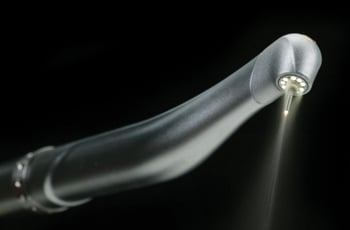
Dr. Meyers of Pinole Periodontics & Dental Implants utilizes the newest in Waterlase MD, which is a revolutionary dental device that uses a combination of laser energy and water to perform many traditional dental procedures without anesthesia or pain.
The Waterlase MD, uses a less invasive approach that conserves healthy tooth structure, which helps teeth function better and last longer.
Waterlase
The Waterlase MD is a revolutionary dental device that uses a combination of laser energy and water to perform many traditional dental procedures without anesthesia or pain. The Waterlase MD, uses a less invasive approach that conserves healthy tooth structure, which helps teeth function better and last longer.
When the traditional dental drill is used, heat and vibration are the major causes of most of the pain. The Waterlase MD works so fast and avoids both heat and vibration, so the tooth nerve can't respond to register pain. This means no shots or the numb feeling after the dental visit.
The Waterlase MD can also perform many soft tissue or gum procedures with little or no discomfort or bleeding.

Pre-Surgery Instructions
Before any oral surgical procedure you should:
- Eat a light and easily digestible meal the night before your appointment
- If you are going to be sedated, DO NOT eat or drink anything on the day of your appointment
- Wear short sleeves and loose-fitting clothing
- Arrange for a relative or friend to stay in the office with you and be ready to drive you home
- You may NOT drive a car on the day of the surgery if you are to be sedated!

Bone Grafting
Bone grafting is commonly performed by an oral and maxillofacial surgeon to replace or augment bone in areas of tooth loss. Bone grafting to the jaws and facial structures may be necessary in a wide variety of scenarios. The most common bone grafts are facial skeleton and jaw procedures. Other common procedures include tooth extraction site graft, bone graft reconstruction and for a sinus lift. Shrinkage of bone often occurs when a tooth is lost due to trauma, severe caries, or periodontal disease. Additionally, bone loss may have already occurred due to infection or pathology around a tooth. There are many artificial biocompatible bone substitutes available; however, the best material for a bone graft is your own bone, which most likely will come from your chin, the back part of your lower jaw or your hip bone. The hip is considered to be a better source because the hip bone has a lot of marrow, which contains bone-forming cells. There are also synthetic materials that can be used for bone grafting. Most bone grafts use a person's own bone, possibly in combination with other materials.
To place the removed bone in the recipient site, little holes are drilled in the existing bone to cause bleeding. This is done because blood provides cells that help the bone heal. The block of bone that was removed will be anchored in place with titanium screws. A mixture of the patient's bone marrow and some other bone-graft material will then be placed around the edges of bone block. Finally, a membrane is placed over the area and the incision closed.
The bone graft will take about 6 to 12 months to heal before dental implants can be placed. At that time, the titanium screws used to anchor the bone block in place will be removed before the implant is placed.
Tooth Extraction
When the extraction of a tooth is required:
- An incision in the gums is made
- The tooth is removed
- The area is stitched up and is allowed to heal
During this time, it is important to think about a tooth replacement option. An extracted tooth leaves an open area in the jaw which, in time, allows the neighboring teeth to drift into the area where the tooth was extracted. This in turn, causes a chain reaction to all the surrounding teeth. Also, if you are considering placing an implant in the future, you should consider asking your dentist to place a bone graft at the time of surgery to preserve the bone width and height.
Wisdom Tooth Problems
The problems involving your wisdom teeth may be caused by the size of your jaw and/or by how crowded your teeth are. Common warning symptoms that there is an un-natural problem in the development of your wisdom teeth could be pain and swelling.
Symptoms can be caused by:
- Infection to the gums
- A crowded tooth displacing neighboring teeth
- A decayed wisdom tooth
- Poorly positioned wisdom tooth
- A cyst that destroys bone
Oral Pathology
The inside of the mouth is normally lined with a special type of skin (mucosa) that is smooth and coral pink in color. Any alteration in this appearance could be a warning sign for a pathological process. The most serious of these is oral cancer.
The following can be signs at the beginning of a pathologic process or cancerous growth:
- Reddish patches (erythroplasia) or whitish patches (leukoplakia) in the mouth.
- A sore that fails to heal and bleeds easily.
- A lump or thickening on the skin lining the inside of the mouth.
- Chronic sore throat or hoarseness.
- Difficulty in chewing or swallowing.
These changes can be detected on the lips, cheeks, palate, and gum tissue around the teeth, tongue, face and/or neck. Pain does not always occur with pathology, and curiously, is not often associated with oral cancer. However, any patient with facial and/or oral pain without an obvious cause or reason may also be at risk for oral cancer. We would recommend performing an oral cancer self-examination monthly and remember that your mouth is one of your body's most important warning systems. Do not ignore suspicious lumps or sores. Please contact us so we may help.

Ankylosis
This refers to a tooth or teeth (primary or permanent) that have become "fused" to the bone, preventing it or them from moving "down" with the bone as the jaws grow. This process can affect any teeth in the mouth, but it is more common on primary first molars and teeth that have suffered trauma (typically the incisors). Treatment can vary depending on the degree of severity of the ankylosis (how "sunken into the gums" a tooth may appear). The degree of severity usually will vary depending on how early the process started, and as a general rule, the earlier it starts, the more severe the ankylosis becomes with age. Several considerations must be taken before any treatment is provided, and your dentist will discuss all the risks and benefits of each treatment option.

Frenectomy
If a patient has an excess amount of tissue that connects the lower and upper lips to the jaw and gum line, a frenectomy procedure is performed to remove the excess tissue. A frenectomy is either performed inside the middle of the upper lip, which is called a labial frenectomy, or under the tongue, called a lingual frenectomy. Frenectomy is a very common dental procedure in the dental world and is performed both on children and adults.
Crown Lengthening
Crown lengthening is a surgical procedure that re-contours the gum tissue and often the underlying bone of a tooth. Crown lengthening is often for a tooth to be fitted with a crown. It provides necessary space between the supporting bone and crown, which prevents the new crown from damaging bone and gum tissue.
Functional Crown Lengthening
Periodontal procedures are available to lay the groundwork for restorative and cosmetic dentistry and/or to improve the esthetics of your gum line. Your teeth may actually be the proper lengths, but they are covered with too much gum tissue. Crown lengthening is a procedure to correct this condition.
During this procedure, excess gum and bone tissue is re-shaped to expose more of the natural tooth. This can be done to one tooth, to even your gum line, or to several teeth to expose a natural, broad smile.
Crown lengthening can make a restorative or cosmetic dental procedure possible. Perhaps your tooth is decayed, broken below the gum line, or has insufficient tooth structure for a restoration, such as a crown or bridge. Crown lengthening adjusts the gum and bone level to expose more of the tooth so it can be restored.
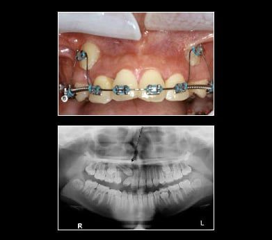
Impacted Canine
Canine teeth are also commonly referred to as cusped or "eye teeth" since the teeth align under your eyes. You should have two canines in both your upper and lower jaw. They are the strongest teeth you have, used for tearing into your most meaty meals. Because of this need for strength, your canines have the longest roots of all your teeth. They are an essential part of your bite and balanced smile for two main reasons:
- Your Bite
Due to their length, the canines guide your other teeth together when chewing and biting. Canines are essential for maintaining a proper bite. - Your Appearance
Without canines, large gaps appear in your smile. This can lead to your other front teeth becoming twisted or crooked.
Your canine teeth are generally some of the last teeth to erupt. Occasionally they do not erupt. The two most common reasons are:
- Overcrowding in your mouth
Extra teeth or a small jaw can cause the space where your canines are supposed to come in to be very small, resulting in impaction, or failure to erupt. - Abnormal growths
Tissue may have developed in your jaw that prevented your canines from reaching the surface.
The fact that teeth don't always come in like they're supposed to highlights the need for regular dental visits when young teeth are developing. If you suspect your child has impacted canines, don't hesitate to make an appointment with Pinole Periodontics & Dental Implants. With regular dental visits, x-rays and examinations, the problem of impacted canines can be found out early when treatment is easier. If you are an adult and your canines have not erupted Pinole Periodontics & Dental Implants can help. Set an appointment today for an x-ray and consultation. Your smile is up there waiting for you.
Treatment for Impacted Canines
After assessing your situation, Pinole Periodontics & Dental Implants will devise a plan to make room for your canines. With a typical oral surgery and the assistance of an orthodontist your canine will find their way into their proper place over time.

Gum Flap Surgery
When deep pockets between teeth and gums (6 millimeters or deeper) are present, it is difficult for a dentist to thoroughly remove the plaque and tartar. Gum flap surgery is a procedure where the gum flap is lifted away from the tooth. Diseased tissue and sometimes bone is removed. The rough surfaces of the tooth are then smoothed by root planing. The area is medicated and the gum flap is replaced and sutured, allowing the bone and gum tissue to heal.
One of the goals of gum flap surgery is to reduce the depth of the periodontal pockets to make them easier to keep clean.
Gum Lift
A gum lift may be performed to create a more even gum line. Patients with a gummy smile can quickly and safely have unwanted tissue removed, thus exposing more tooth to shape a more attractive smile.
What is a Laser Gingivectomy?
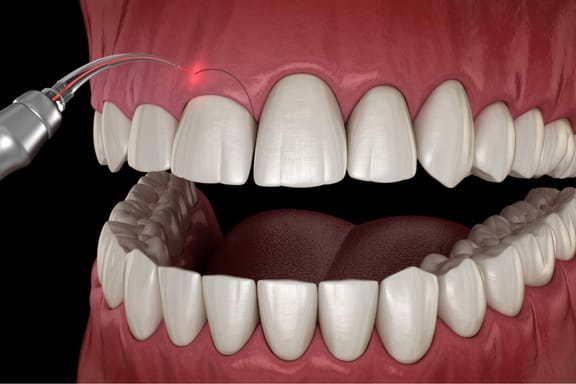
A laser gingivectomy is a cosmetic and periodontal procedure that uses a laser to remove excess or overgrown gum tissue. It is performed to improve the appearance of the gums and enhance the aesthetics of the smile. The procedure is used to correct a "gummy" smile, where the gums appear too prominent, or to treat gum disease, which can cause the gums to recede and expose the roots of the teeth.
Laser gingivectomy differs from traditional gingivectomy, which involves the surgical removal of gum tissue with a scalpel or other surgical instrument. Laser gingivectomy is a minimally invasive procedure that uses a highly precise laser to remove only the targeted tissue. This results in less bleeding, swelling, and discomfort for the patient, as well as a faster and more predictable recovery.
Additionally, the laser cauterizes the tissue as it is removed, reducing the risk of infection and the need for sutures. The laser also seals the blood vessels, reducing bleeding and promoting faster healing. The laser's precision and ability to cauterize the tissue also helps to minimize the damage to surrounding healthy tissue, resulting in a more aesthetically pleasing outcome.

Laser Pocket Disinfection (LPD)
- Kills bacteria and disrupts biofilm to reduce inflammation
- No shots needed
- Maintain healthy gums and avoid progression of gum disease
- No antibiotics needed, avoids antibiotic resistance
- The laser light can reach and destroy bacteria up to 6mm beyond the surface to help prevent bacteria from spreading back into the pocket
- Safe for medically compromised patients, those on blood thinners, or those with diabetes

Frequently Asked Questions About LPD
By removing the inflammation, your body has the energy to heal naturally. The laser light kills bacteria and creates a clean environment for healing.
If you have gingivitis, LPD can help reverse symptoms. LPD can also be used to treat patients on a perio-maintenance program.
Not at all! LPD is a quick and painless. If you are very sensitive, a topical anesthetic may be used.
There are no known side-effects of LPD in over 25 years of therapy, just bigger smiles. The treatment is safe for use around crowns, bridges, sealants, and implants.
LPD is beneficial to patients who have recently had surgery, or are scheduled to have surgery to reduce bacteria that may enter the bloodstream.
Gum disease has been linked to serious health concerns including heart disease, stroke, diabetes, pre-term birth, certain cancers & more.

Post-Surgery Instructions
Fold a piece of clean gauze into a pad thick enough to bite on and place directly on the extraction site. Apply moderate pressure by closing the teeth firmly over the pad. Maintain this pressure for about 30 minutes. If the pad becomes soaked, replace it with a clean one as necessary. Do not suck on the extraction site (as with a straw). A slight amount of blood may leak at the extraction site until a clot forms. However, if heavy bleeding continues, call your dentist. (Remember, though, that a lot of saliva and a little blood can look like a lot of bleeding.)
The Blood Clot
After an extraction, a blood clot forms in the tooth socket. This clot is an important part of the normal healing process. You should therefore avoid activities that might disturb the clot.
Here's how to protect it:
- Do not smoke, rinse your mouth vigorously or drink through a straw for 24 hours.
- Do not clean the teeth next to the healing tooth socket for the rest of the day. You should, however, brush and floss your other teeth thoroughly. Gently rinse your mouth afterwards.
- Limit strenuous activity for 24 hours after the extraction. This will reduce bleeding and help the blood clot to form. Get plenty of rest.
- If you have sutures, your dentist will instruct you when to return to have them removed.
Medication
Your dentist may prescribe medication to control pain and prevent infection. Use it only as directed. If the medication prescribed does not seem to work for you, do not increase the dosage. Please call your dentist immediately if you have prolonged or severe pain, swelling, bleeding, or fever.
Swelling & Pain
After a tooth is removed, you may have some discomfort and notice some swelling. You can help reduce swelling and pain by applying cold compresses to the face. An ice bag or cold, moist cloth can be used periodically. Ice should be used only for the first day. Apply heat tomorrow if needed. Be sure to follow your doctor's instructions.
Diet
After the extraction, drink lots of liquids and eat soft, nutritious foods. Avoid alcoholic beverages and hot liquids. Begin eating solid foods the next day or as soon as you can chew comfortably. For about two days, try to chew food on the side opposite the extraction site. If you are troubled by nausea and vomiting, call your dentist for advice.
Rinsing
The day after the extraction, gently rinse your mouth with warm salt water (teaspoon of salt in an 8 oz. glass of warm water). Rinsing after meals is important to keep food particles away from the extraction site. Do not rinse vigorously!
